
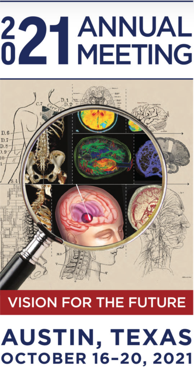
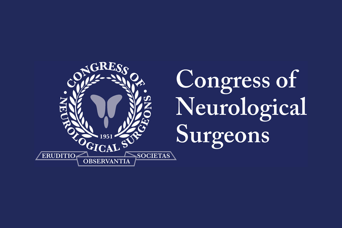
Durante el último congreso americano de Neurocirugía CNS, realizado en Austin, Texas, Oct. 2021, el Dr. Mario Izurieta participó como profesor invitado en la conferencia sobre el manejo de Trauma de Cráneo en el Ecuador y en la mesa redonda sobre neuralgia del Trigémino, junto a otras personalidades de la neurocirugía mundial para discutir sobre el mejor tratamiento.
neurocirugia especializada
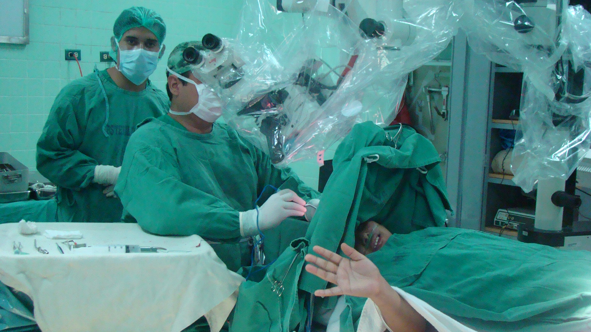
de alta complejidad
MIEMBRO DE ASOCIACION AMERICANA DE NEUROCIRUGIA
PROFESOR INVITADO

Ashok R. ASthagiri, MD
Scientific Program Chair
“El comité científico lo ha seleccionado a Ud debido a su experiencia y reputación sobre la neuralgia del trigémino…”
Ashok R. Asthagiri, MD
Servicios
que tratamientos ofrecemos
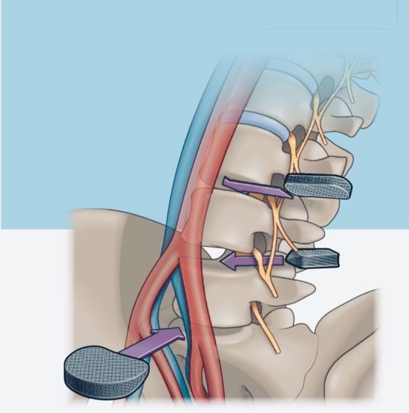
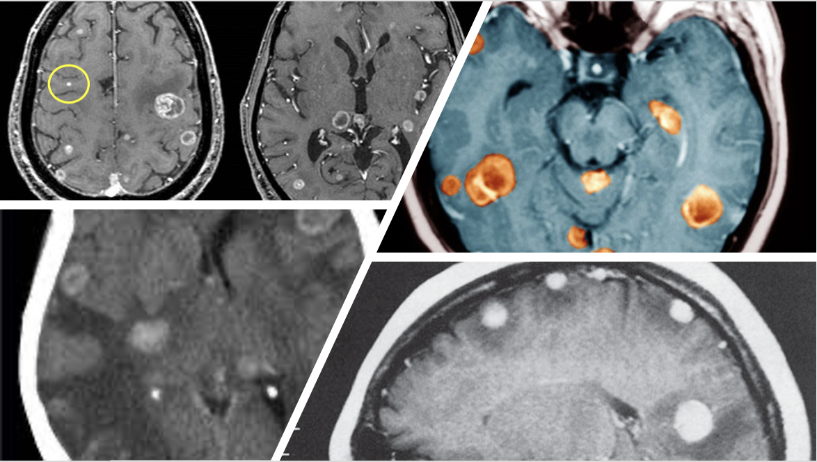
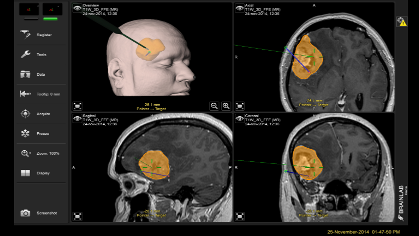
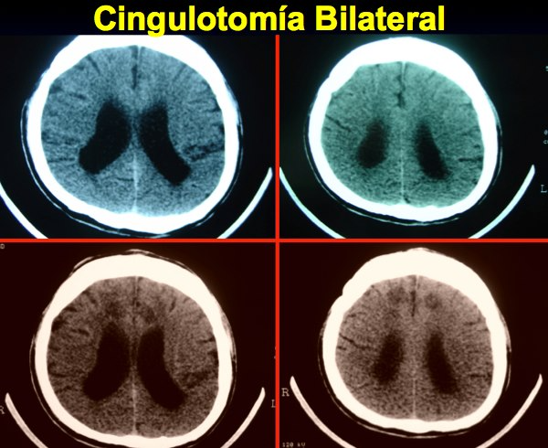
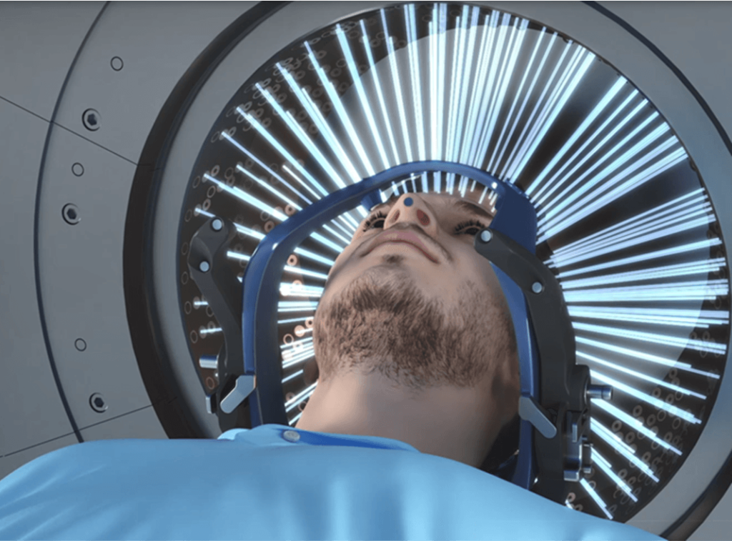
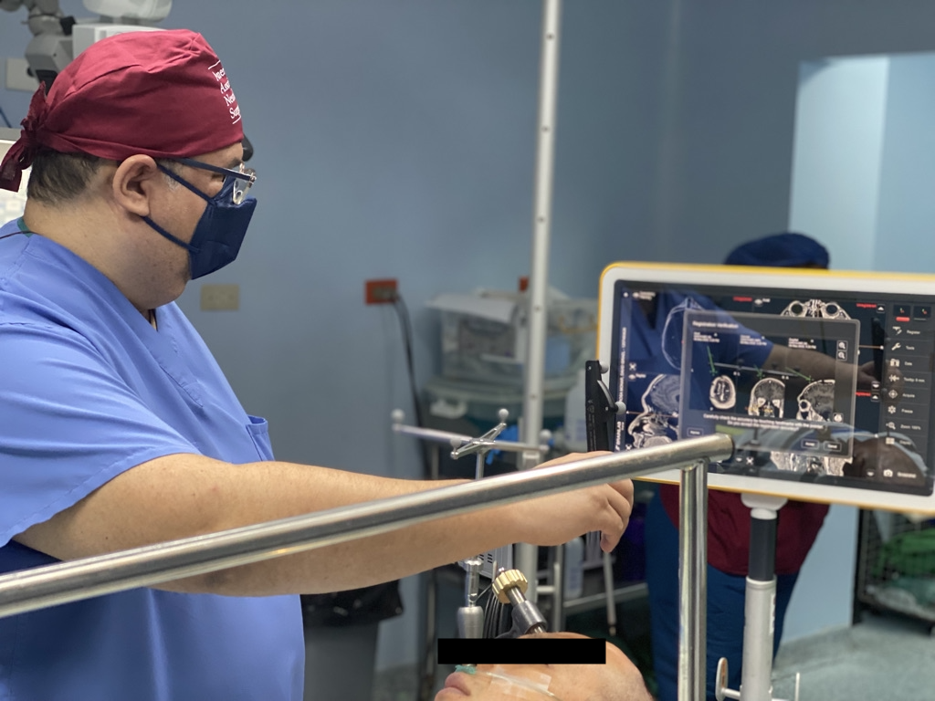
Por medio de instrumentos y sistema de alta tecnología, en la actualidad podemos ofrecer tratamiento computarizado de tumores, epilepsia intratable…
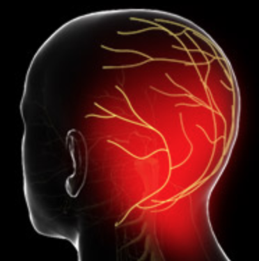
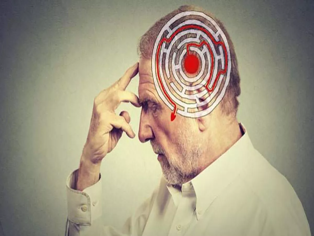
La cefalea mas invisible
CURRICULUM VITAE
ESTUDIOS, PRESENTACIONES Y PARTICIPACION INTERNACIONAL
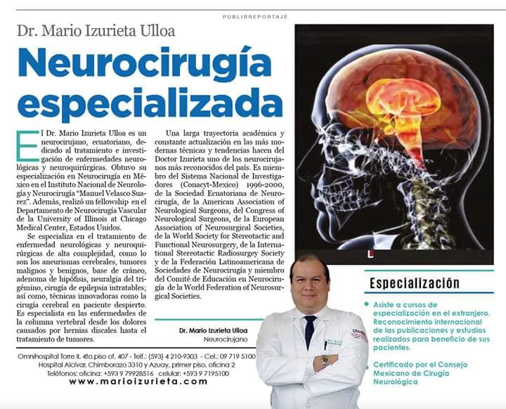
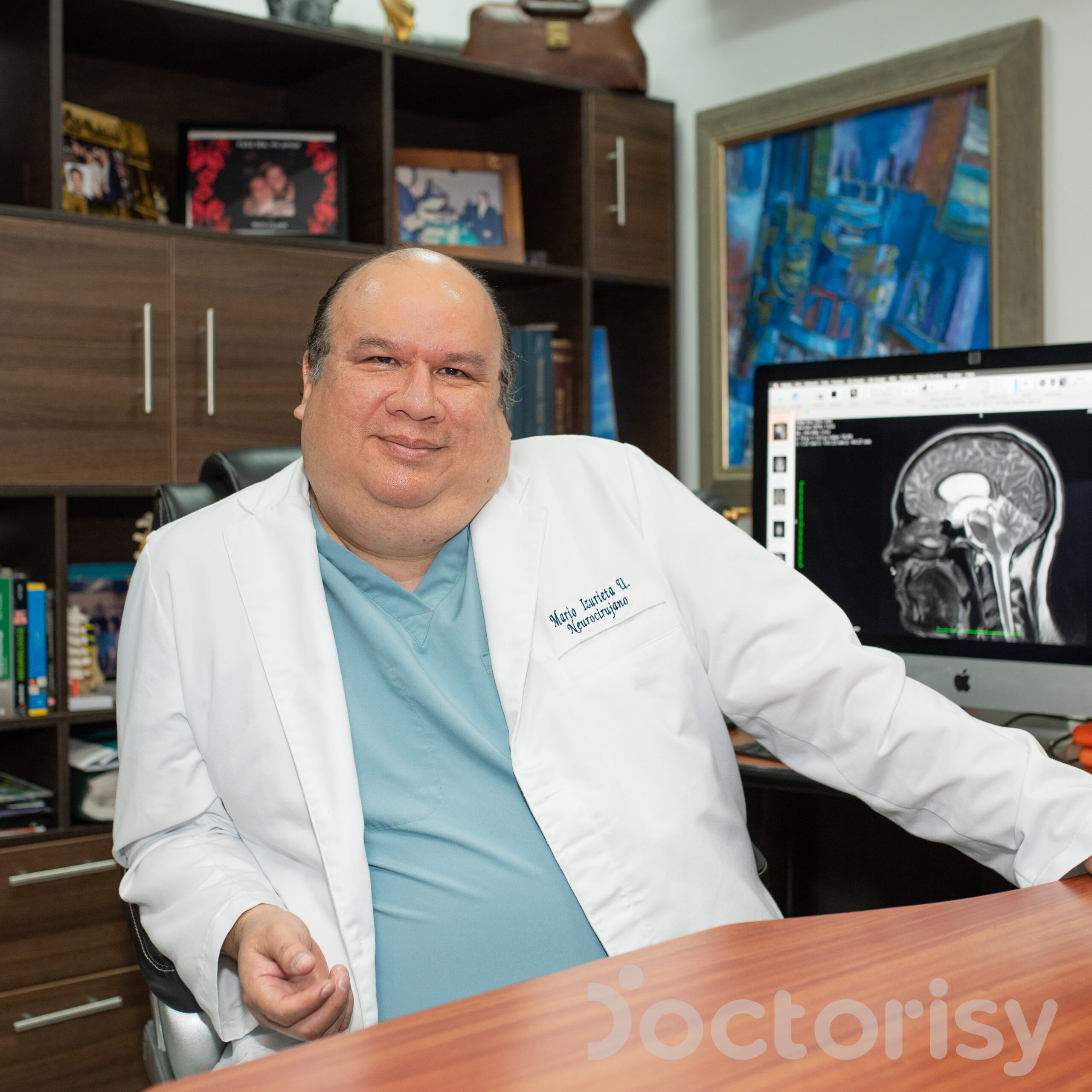
El Dr. Mario Izurieta Ulloa es un neurocirujano, ecuatoriano, dedicado al tratamiento e investigación de enfermedades neurológicas y neuroquirúrgicas. Obtuvo su especialización de Post-grado en Neurocirugía en Mexico, en el Instituto Nacional de Neurología y Neurocirugía «Manuel Velasco Suarez». Además, realizó un fellowship en el Departamento de Neurocirugía Vascular de la Universidad de Illinois en Chicago (UIC)…
INVESTICAGION MEDICA
30 AÑOS DE EXPERIENCIA EN INVESTIGACION MEDICA
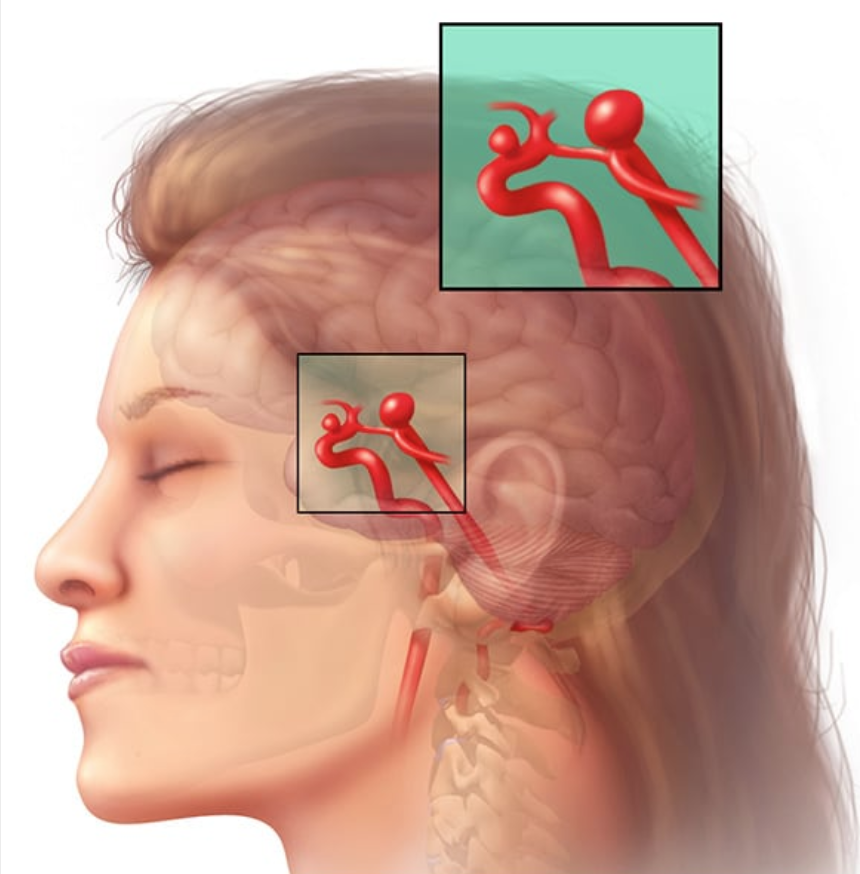
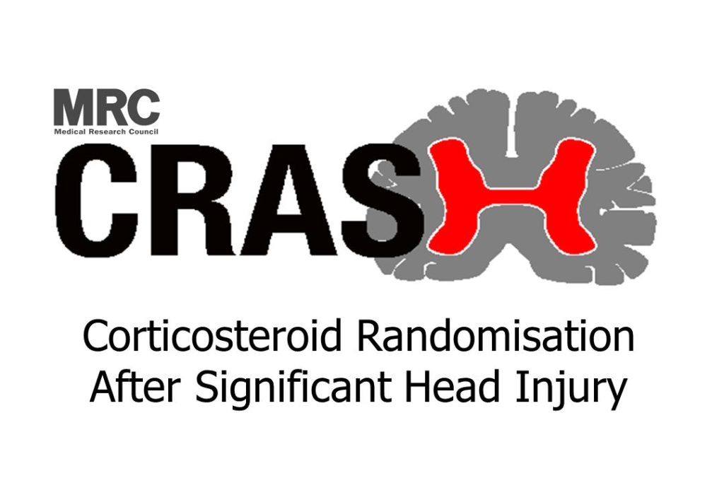
El Dr. Mario Izurieta ha liderado varios estudios a nivel nacional con participación internacional como son el grupo de estudios CRASH, organizado por la famosa Escuela de Medicina e higiene Tropical de Londres, Inglaterra y publicados en la revistas médicas mas importantes del mundo.


Desea un consulta médica? Presencial o Virtual?
Estamos encantados de recibirlos por medio de una consulta presencial o virtual por medio de medios digitales disponibles.
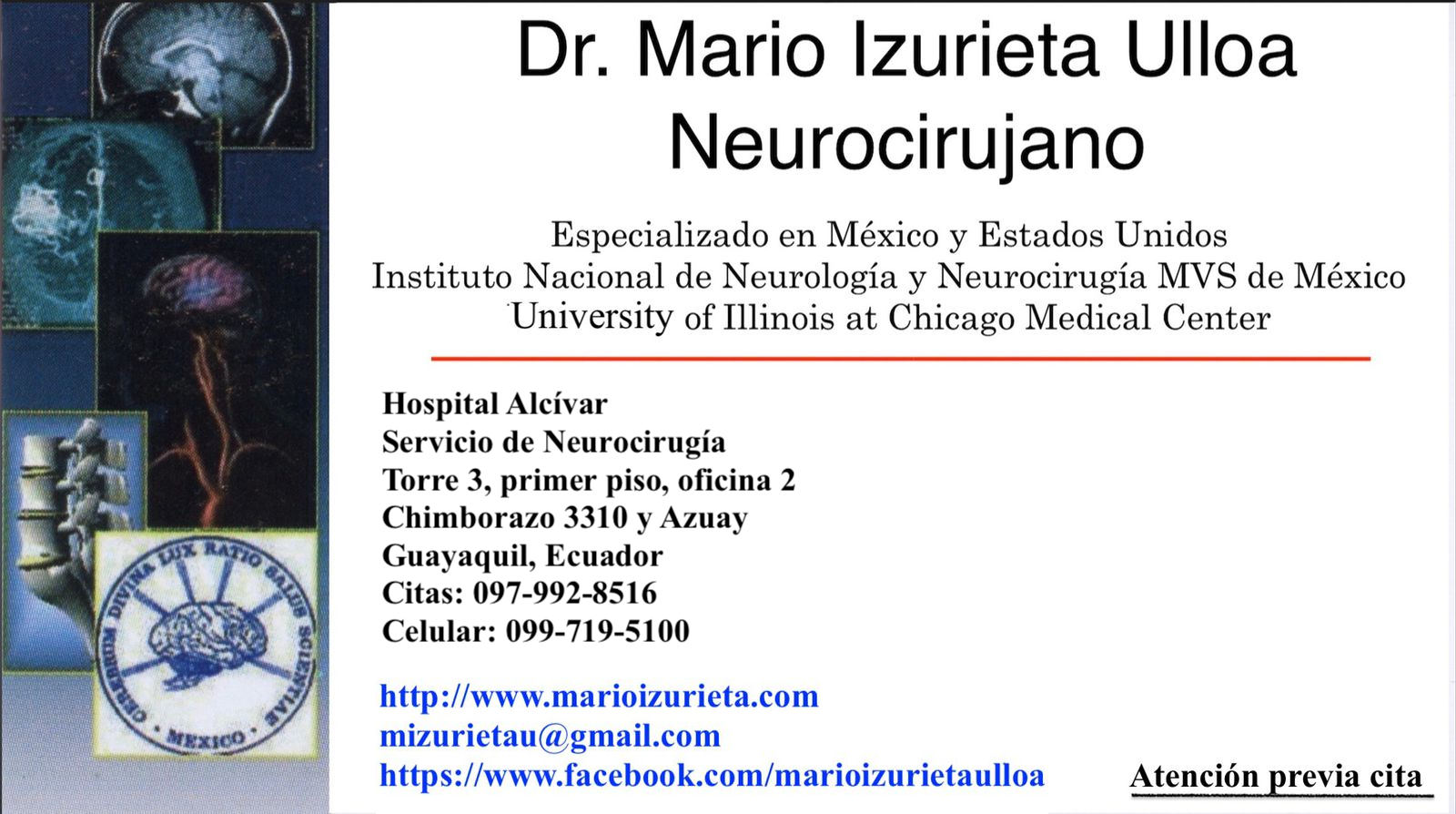
Clínica Alcívar Torre 3, 1er Piso, ofc.2
Chimborazo 3310 y Azuay
Teléfonos: 097 992 8516 / 099 719 5100
Otras Páginas
Links Rápidos
Ultimos Proyectos
marioizurieta.com webpage design MIU
Copyright © 2023. All rights reserved.